Printable Worksheet of The Circulatory System
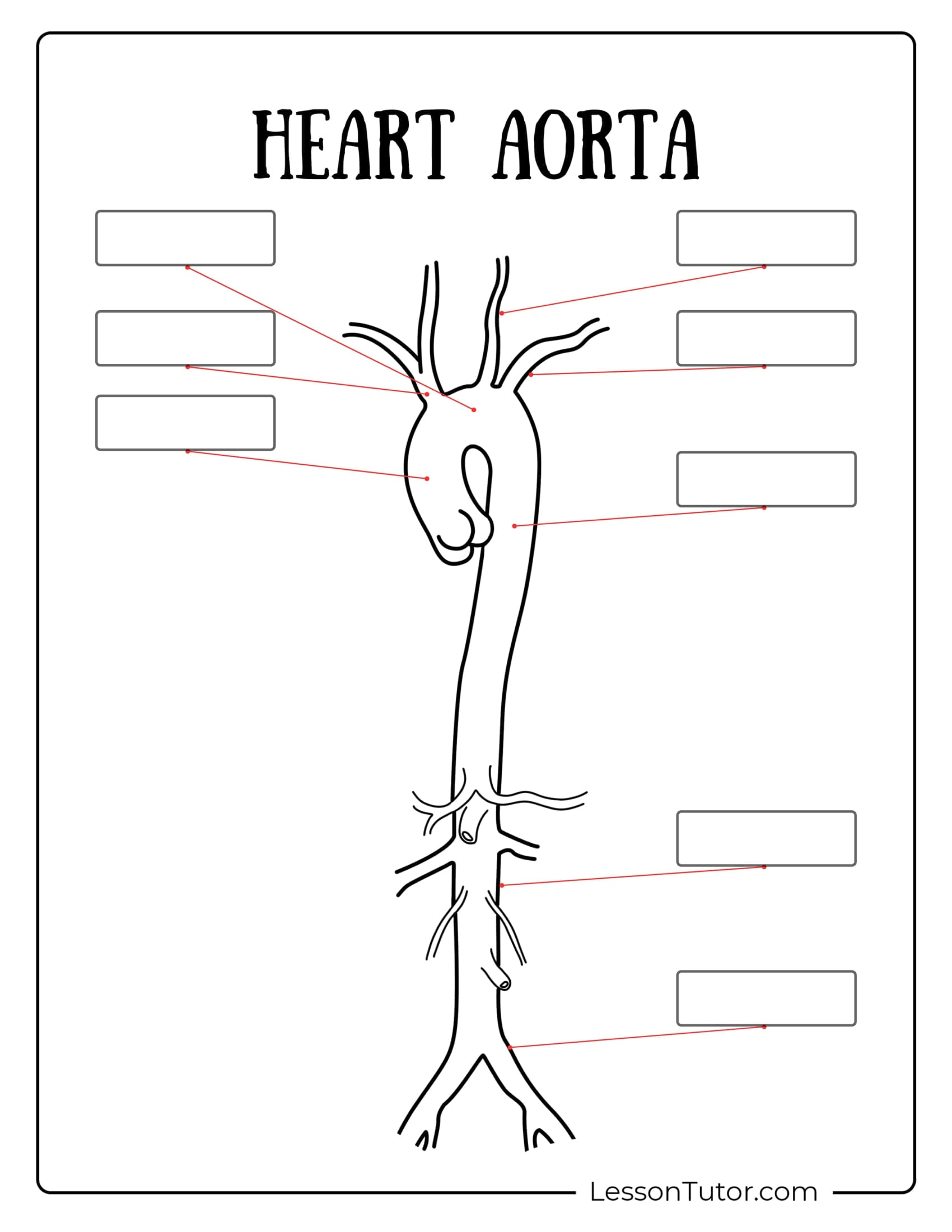
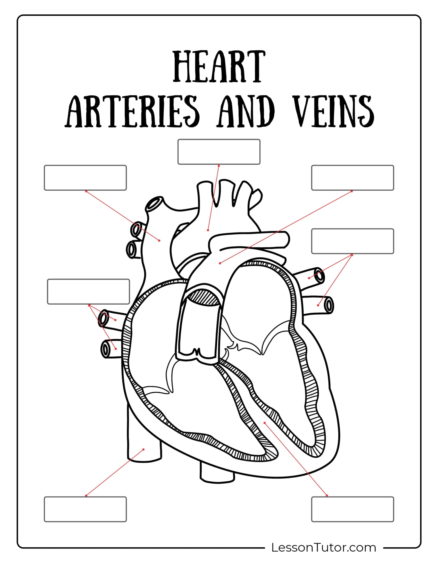
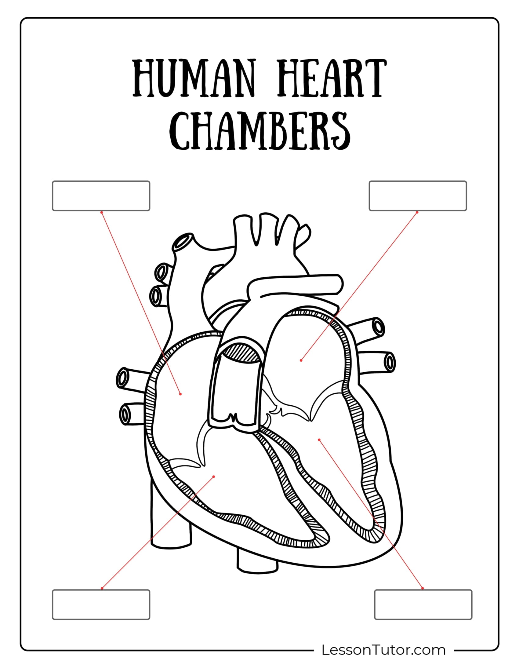
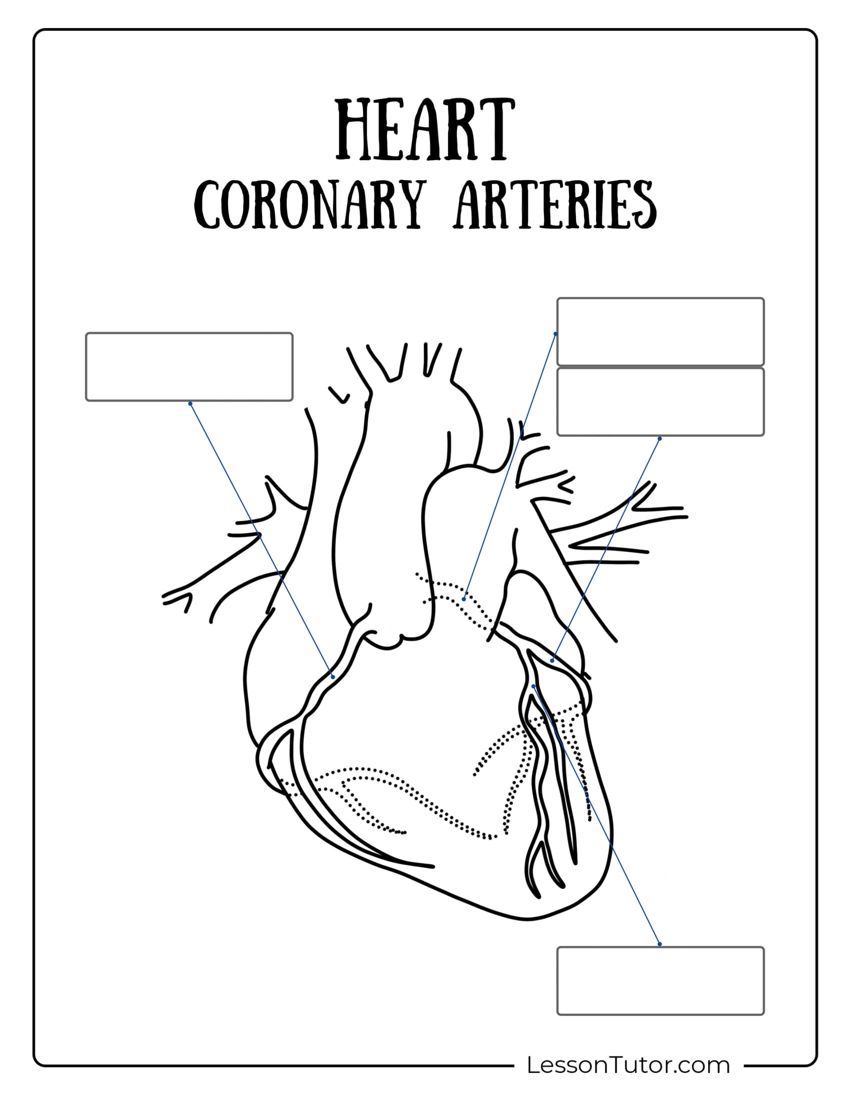
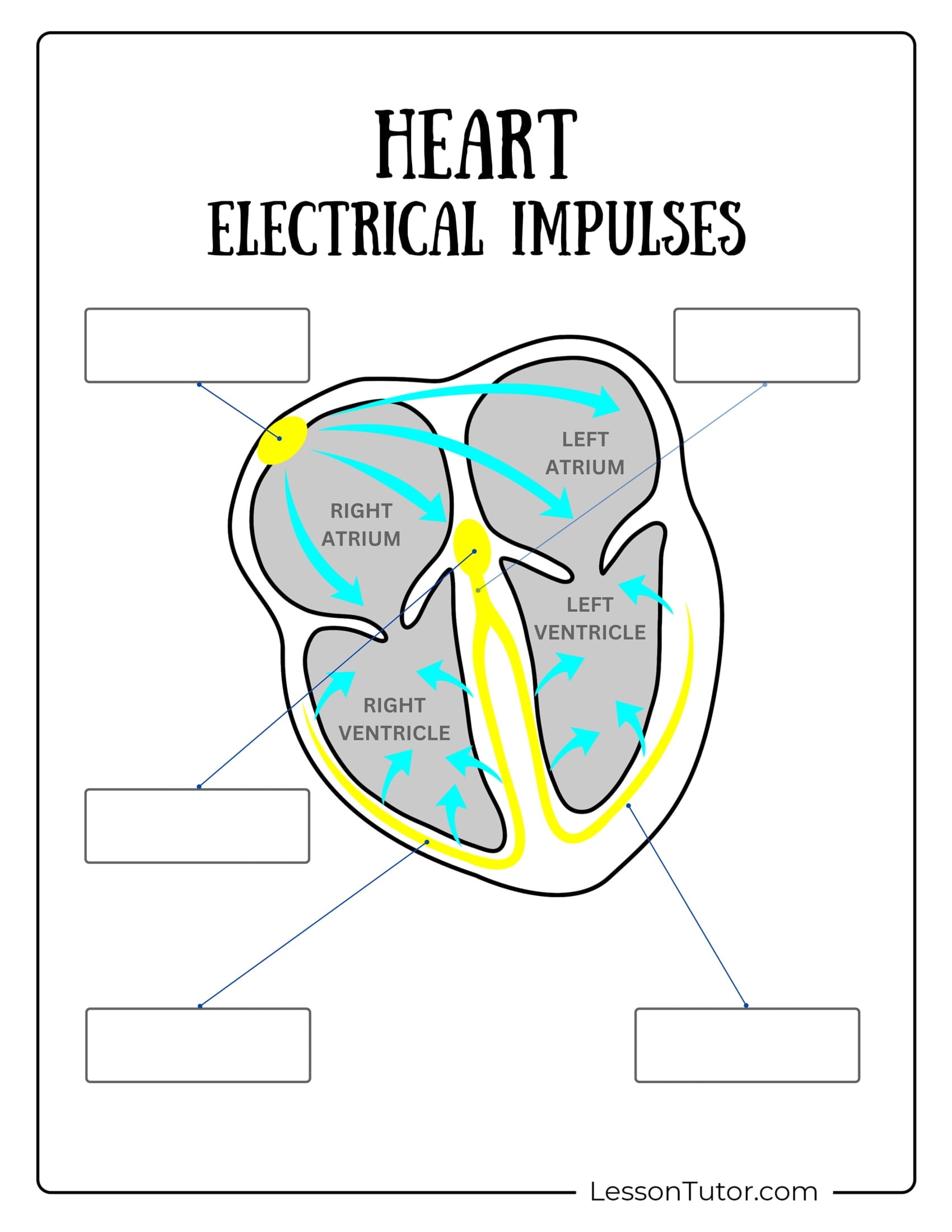
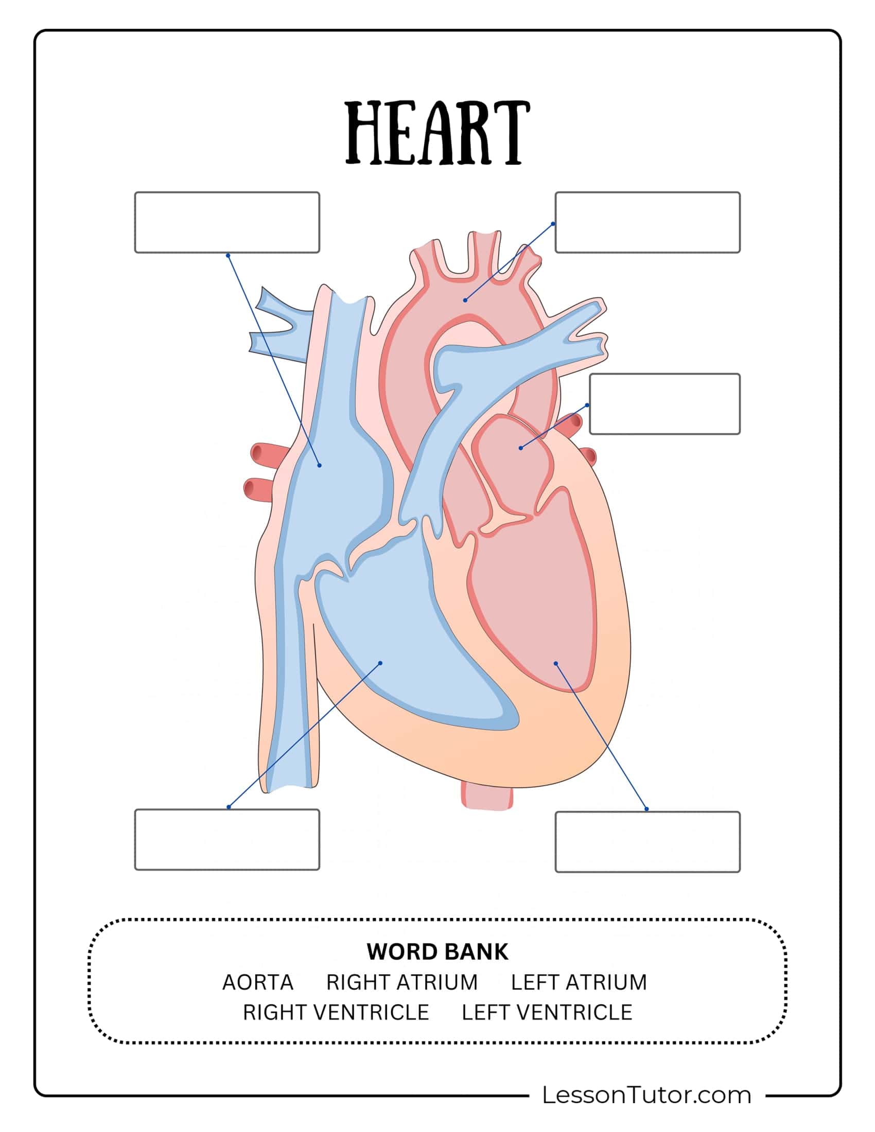
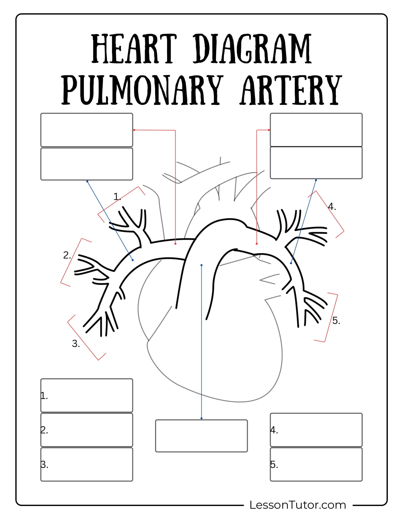
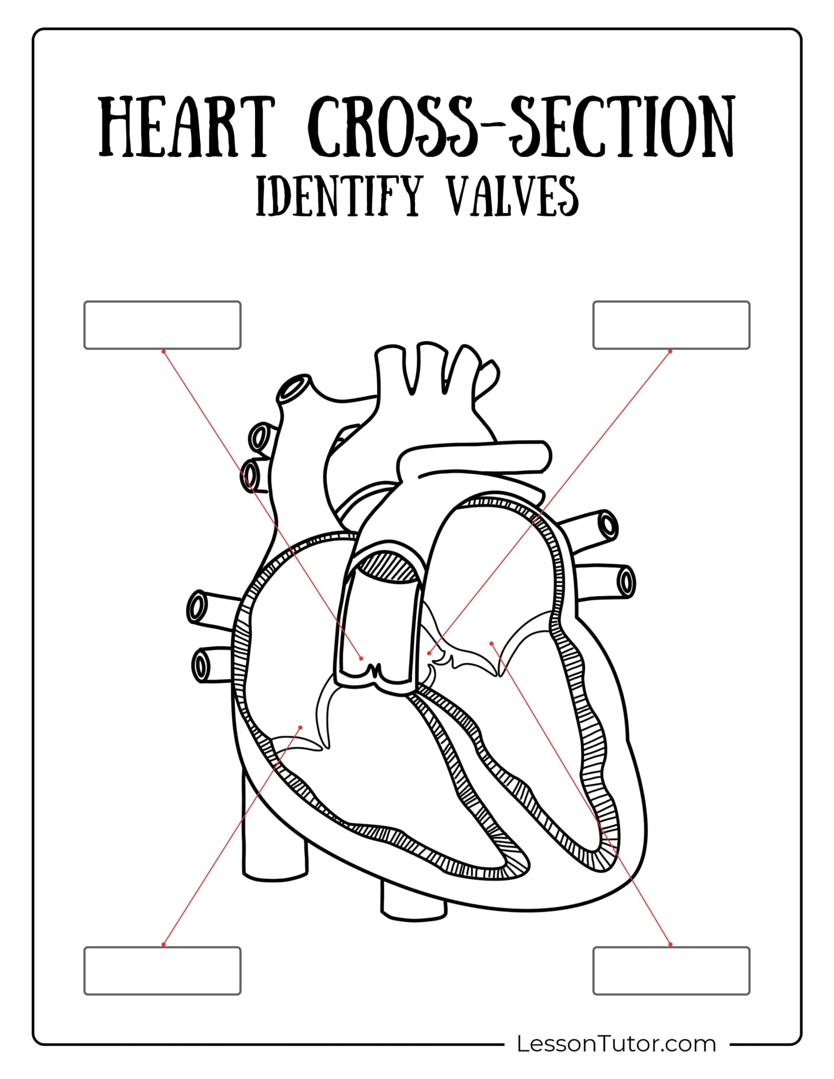
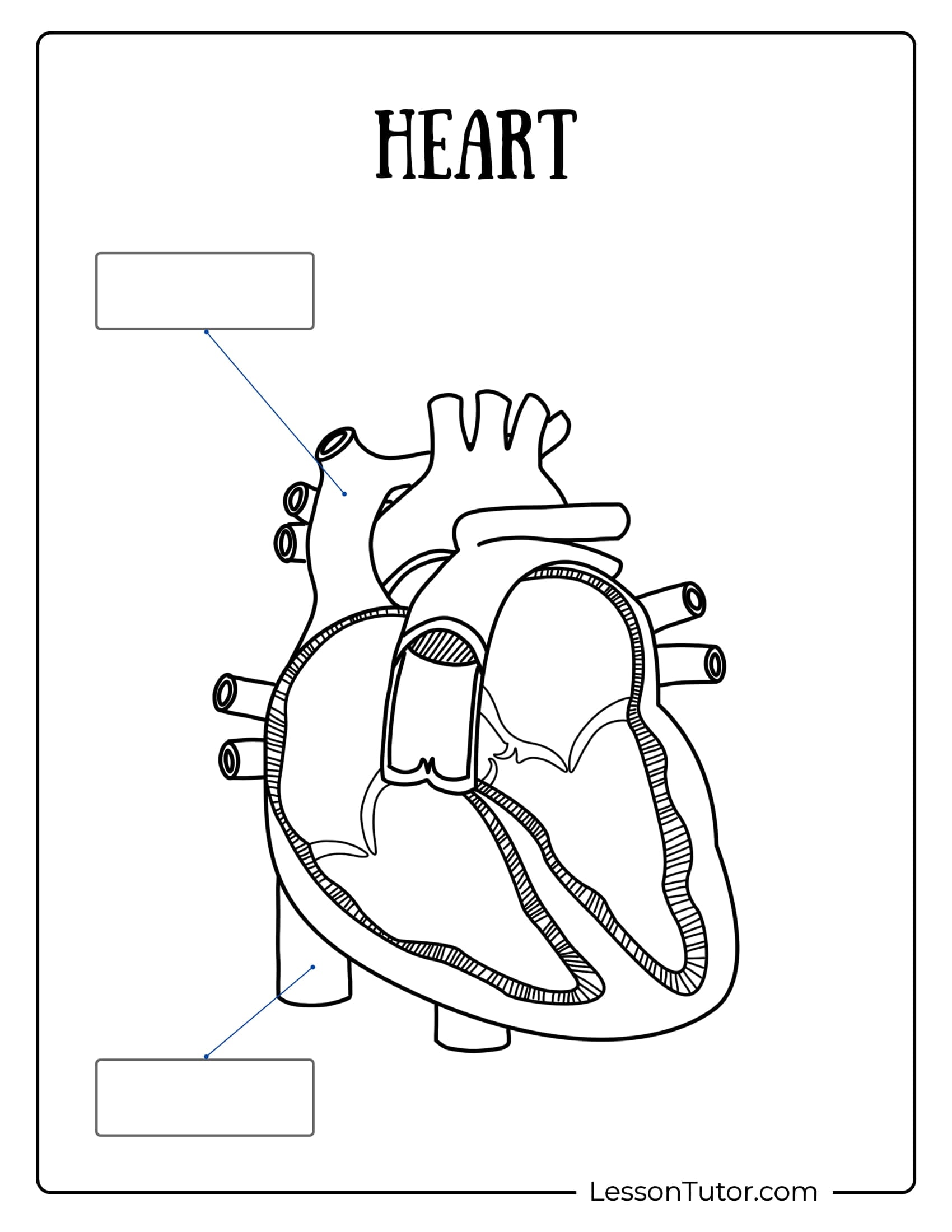
(10) I. Label the diagram above:
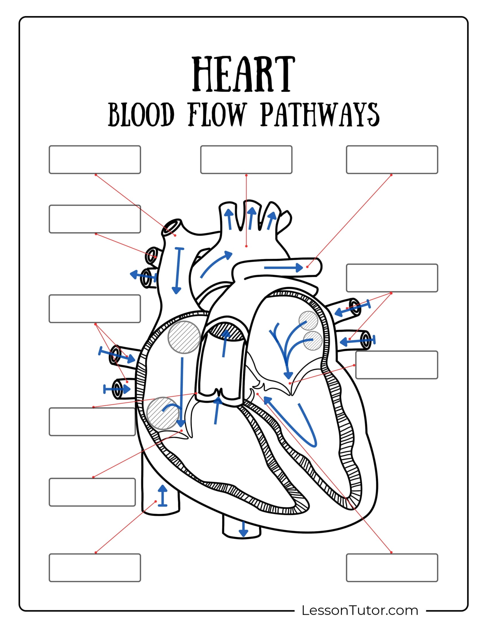
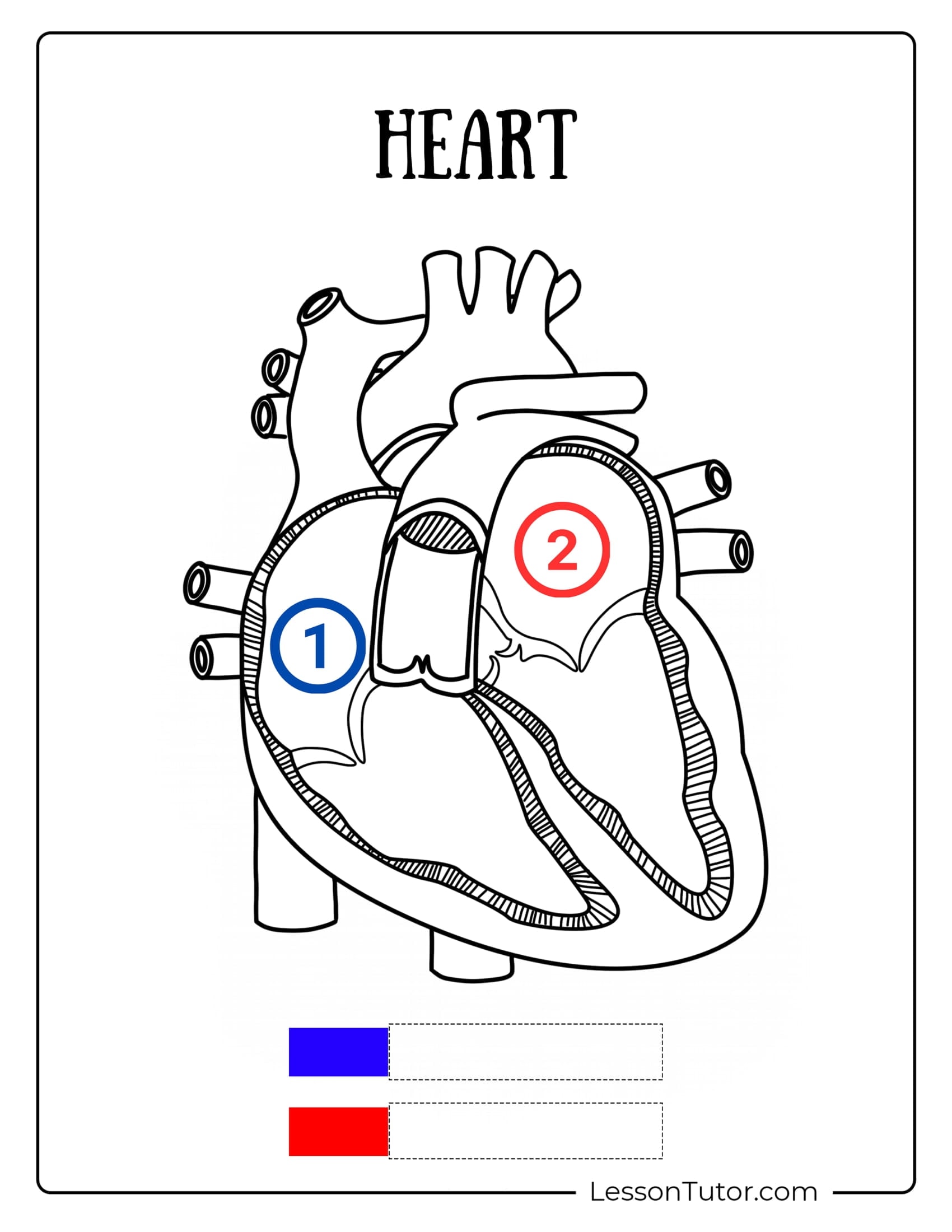
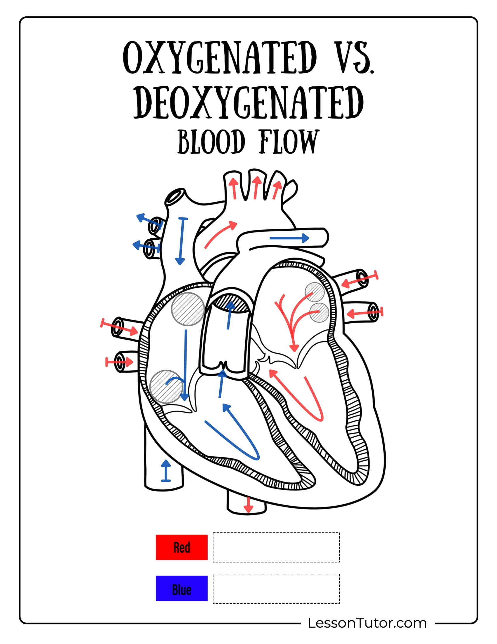
(4) II. Colour the path of oxygenated blood red and deoxygenated blood blue. (6) III. Use arrows to indicate blood flow direction. (1) IV. Capillaries are to body tissues as _______________ are to lungs. (4) V. Match the organ with its corresponding blood supply:
|
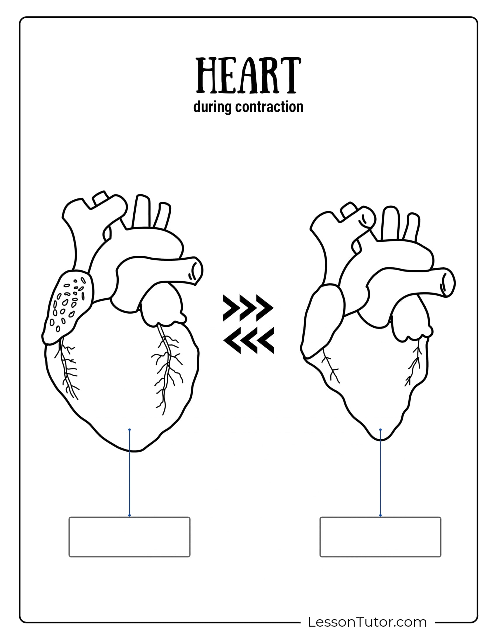
Every structure in the body needs blood to supply it with oxygen and nutrients. These same structures also need a means to get rid of their own waste products. Very simply, each organ has a one-way pipe going into it, feeding it, and a one way pipe going out, like flushing a toilet.So – where does all this start? If you study the simplified diagram, right, you’ll see that the blood travels endlessly around in a modified figure-8. Blood is pumped by the heart’s contractions. Without a beating heart, there would be no blood flowing anywhere. There is a reason why this diagrams of the circulatory system shows a ‘flattened’ version of the circulatory system. It is so much easier to see the route of the blood around and around the body this way, rather than a 3-D reproduction of how the blood vessels actually twist and loop and circle other impeding structures.(see below) This simplified version also distinctly shows that the lungs are treated differently from the rest of the body. They are the exception to a number of circulatory ‘rules.’ (If they were the same, the pictures would be of an endless circle) More on this later. R.A. = Right Auricle or Atrium 1 = Pulmonary Vein |
Blue Blood or Red Blood?
Rule #1: All Arteries flow Away from the heart.
There are many, many organs in the body. Each has its own artery. For some, it is easy to identify the organ for which they are intended – like the pancreatic arteryfeeds the pancreas. Others are a little more challenging to remember:
Pulmonary artery -> lungs
gastric artery -> stomach
hepatic artery -> liver
renal artery -> kidney
coronary artery -> heart
carotid artery -> brain
Aorta -> Almost all arteries branch from this major blood highway (exception = pulmonary artery)
Rule #2: Veins always lead to the heart.
Once the artery reaches its target destination, you will notice that it divides and subdivides, like the branches of a tree. This is so that the oxygen rich blood is undiluted before it reaches every nook and cranny of the receptive organ. Arteries divide into arterioles which divide into capillaries. The fascinating transfer work happens here in the capillaries. Imagine your right hand is the capillary end of an arteriole (your right arm). Lace your 2 hands together. Your left hand represents the capillary end of a Vein. Here is where the oxygen is discharged and the empty hemoglobin containers are recycled back towards the heart on their path to re-filling. Once the oxygen portion is used, the blood tends to lose its bright red colour. Venous blood (blood found in veins) tends to be darker in appearance. It is also more sluggish in speed as it is now so far away from the heart, and using your imagination and the diagram above, it also has to travel ‘up-hill.’ Veins are lined with little valves, or shelves, that help this slow stuff from sliding ‘backwards’ along the road back to the heart. Blood from an artery would literally spurt to a beat if it were inadvertently opened. Most of these ‘V’ words (veins, venules, venous, vena…) all stem from the Latin verb “venire,” meaning ‘to come.’
Rule #3: All Arteries carry Oxygenated Blood EXCEPT the Pulmonary Artery
2 Major Arteries: Pulmonary Artery and the Aorta
The Pulmonary Artery carries blood away from the heart and to the lungs. Once it arrives, fresh, oxygenated blood plumps up the hemoglobin, like filling a gas tank at a gas station pump. Now it continues back to the heart’s clover-leaf for distribution first to the coronary arteries and then on to the rest of the body.
Oxygenated blood flows out of the heart via the Aorta – the Mother of all arteries and also the largest. When you see a diagram of the circulatory system you’ll note that on exiting the Left Ventricle, this blood vessel is called the Ascending Aorta. The next portion, giving rise to multiple branches, is called the Arch (not unlike the golden McDonald’s arches, only upside down). These tributaries are: a) the Carotid artery, which supplies blood to the head and neck. b) the Subclavian arteries, which supply blood to the upper chest and arms, c) the Coronary arteries, which supply the heart itself with oxygen rich blood. Once past these exits, the Aorta dives down in direction and is aptly named the Descending Aorta.
Rule #4 All Veins carry de-oxygenated blood EXCEPT the Pulmonary Vein
Added Details
Excerpt from: http://www.hearttalk.net/content/ah03.htm
“…4. In the tiny capillaries, each so narrow that red blood cells must pass through one at a time, the actual exchange of nutrients and waste products takes place at the cellular level.
5. The blood, now oxygen poor, travels through the capillaries to small veins called venules, and then to larger veins, such as the saphenous veins in the legs and hips. Veins in the lower part of the body connect with the inferior vena cava, and veins in the upper part of the body connect with the superior vena cava. Both of these venae cavae empty into the upper right chamber (right atrium) of the heart.
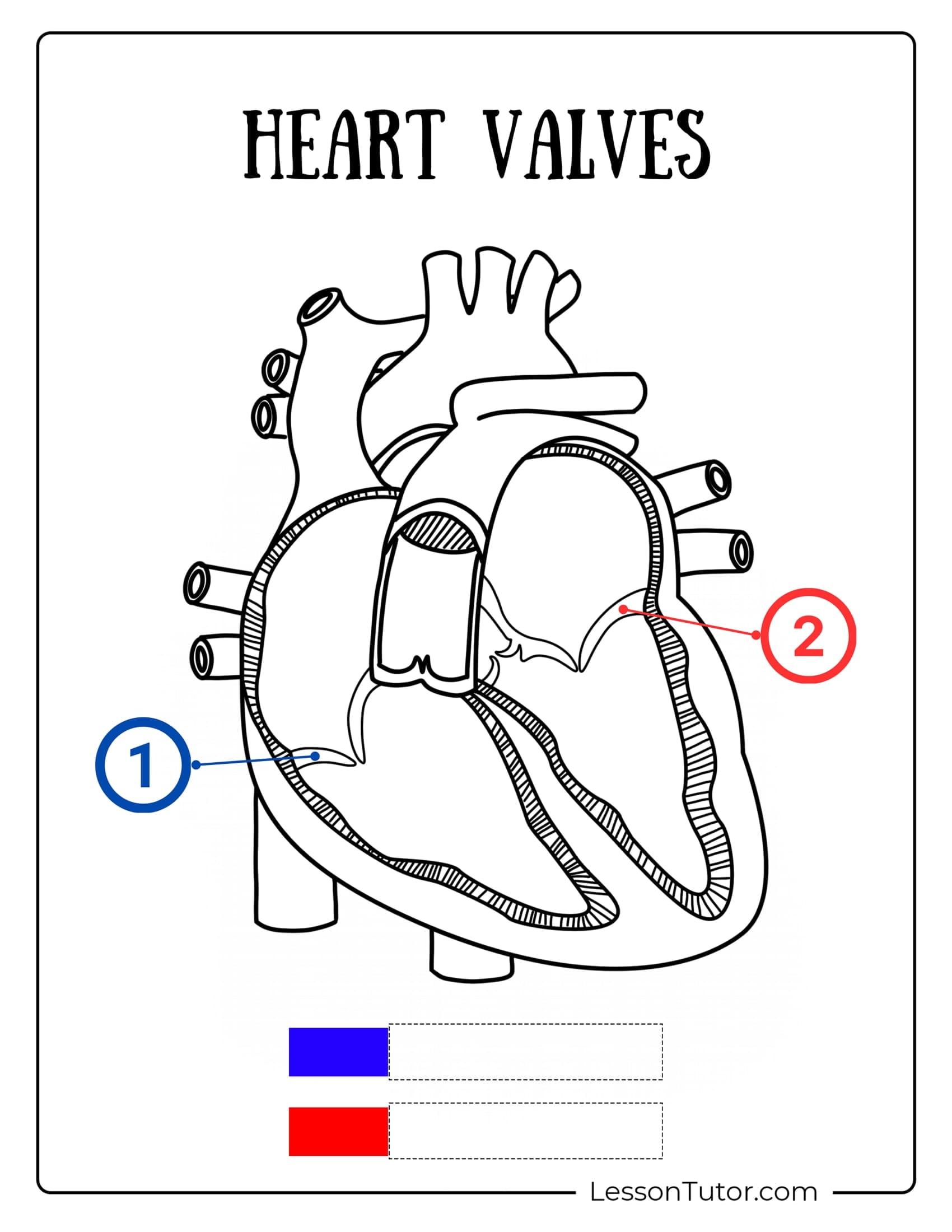
6. When the right atrium is filled, the tricuspid valve opens to allow blood to fill the right ventricle. When signaled by an electrical impulse, the ventricle pumps the blood through the pulmonic valve and out the pulmonary artery to the lungs. The oxygen poor blood flows into air sacs in the lungs (called alveoli), where an exchange of gases is completed. The oxygen poor blood gives up its carbon dioxide waste (which is exhaled from the body) in exchange for new molecules of oxygen. As the blood absorbs the fresh oxygen, it once again turns a healthy red color.
7. Oxygen rich blood returns from the lungs to the heart via the pulmonary veins, emptying into the left atrium. When the left atrium is filled, the mitral valve opens to allow blood to fill the left ventricle. When signaled by an electrical impulse, the left ventricle pumps the oxygen rich blood through the aortic valve and out the aorta, beginning the cycle all over again. Some oxygen rich blood also returns to the heart via the coronary arteries, as described in the next section….”
More Articles by this Consultant, Click Here
More Articles in this Subject, Grade 5 Biology, Click Here
More Articles for General Science, Grade 5, Click Here.